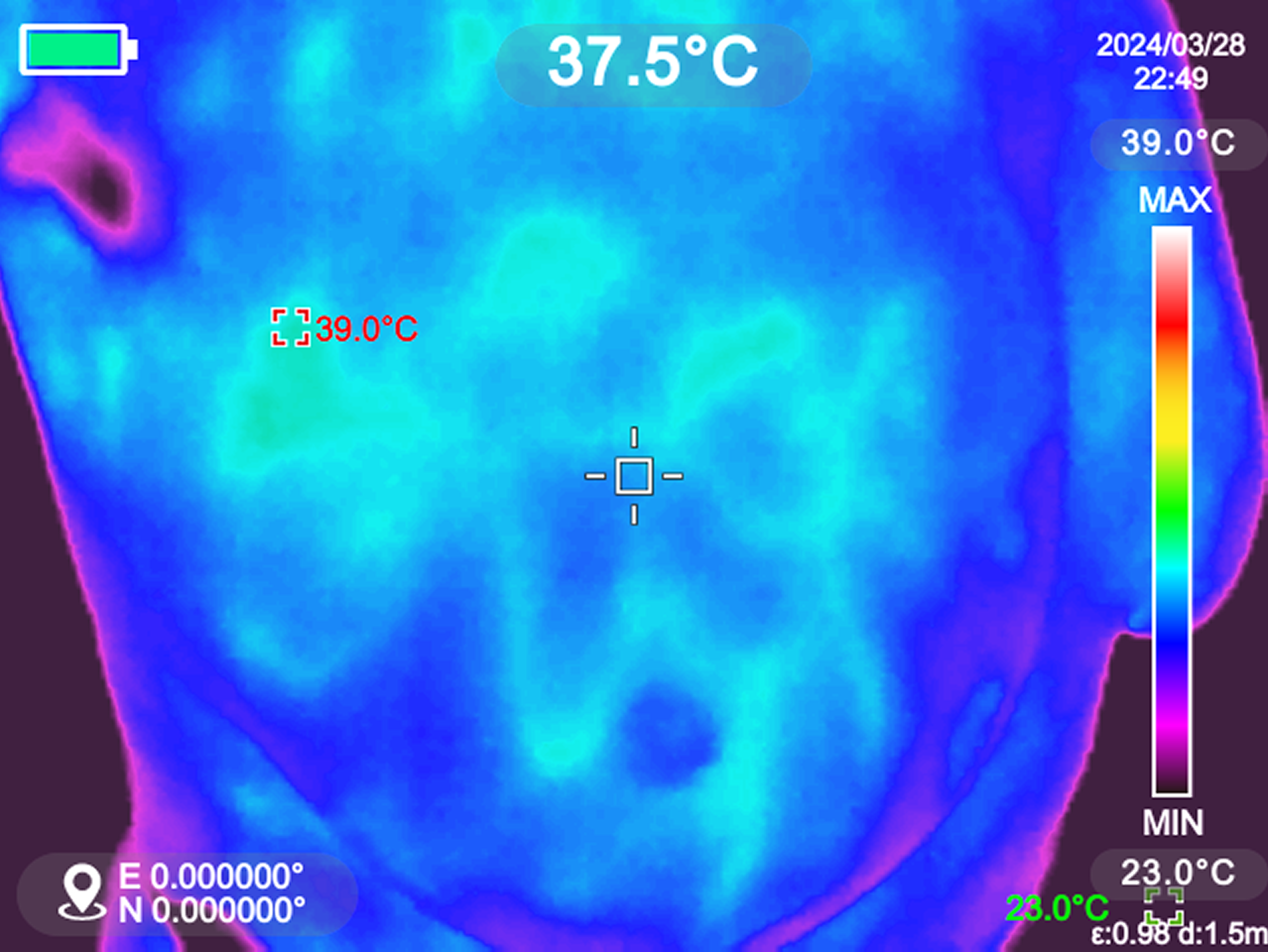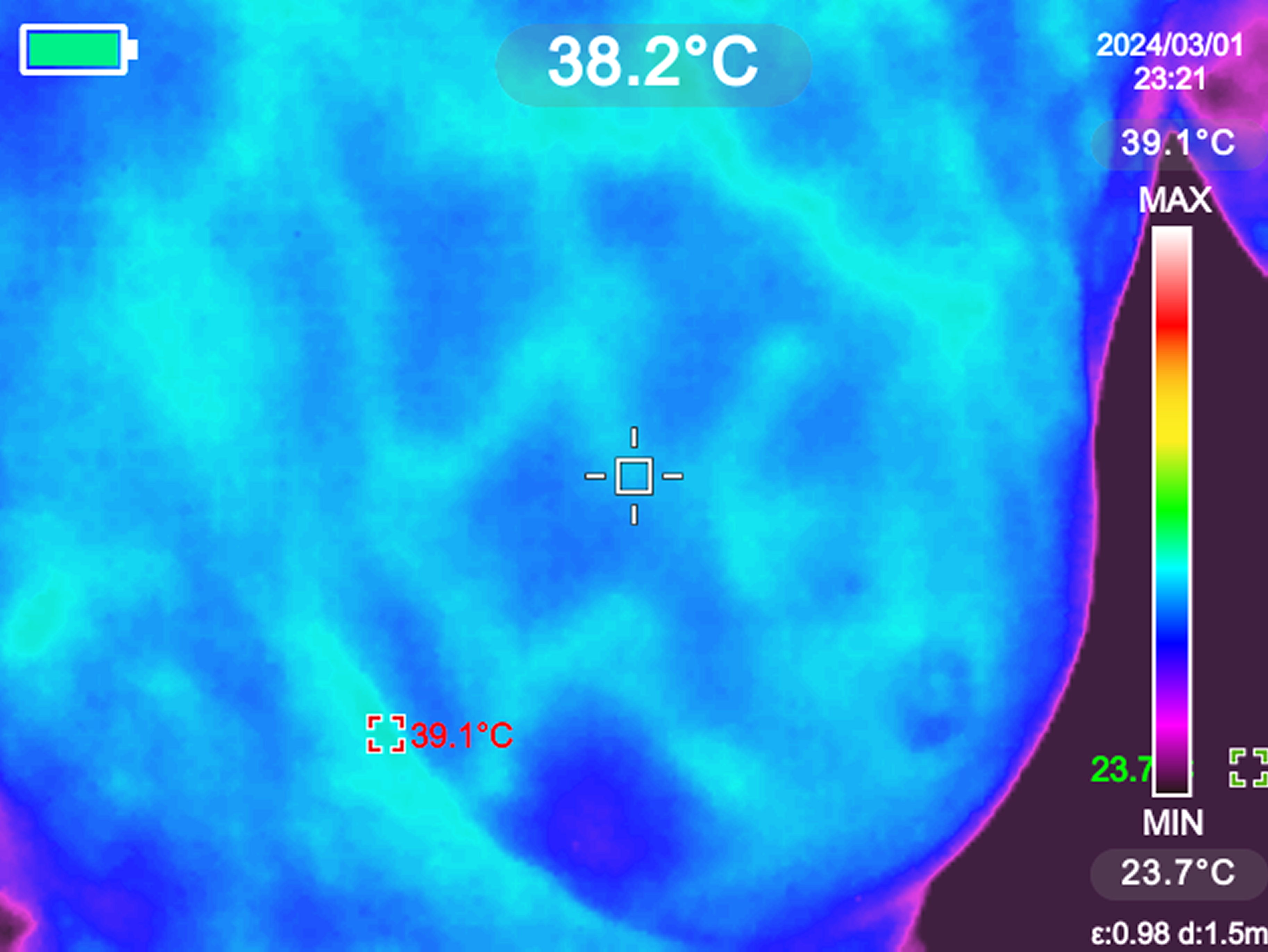
Thermography
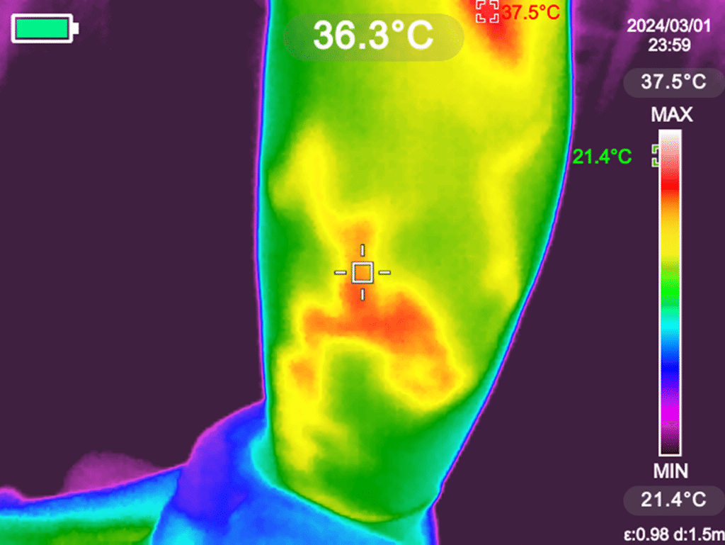
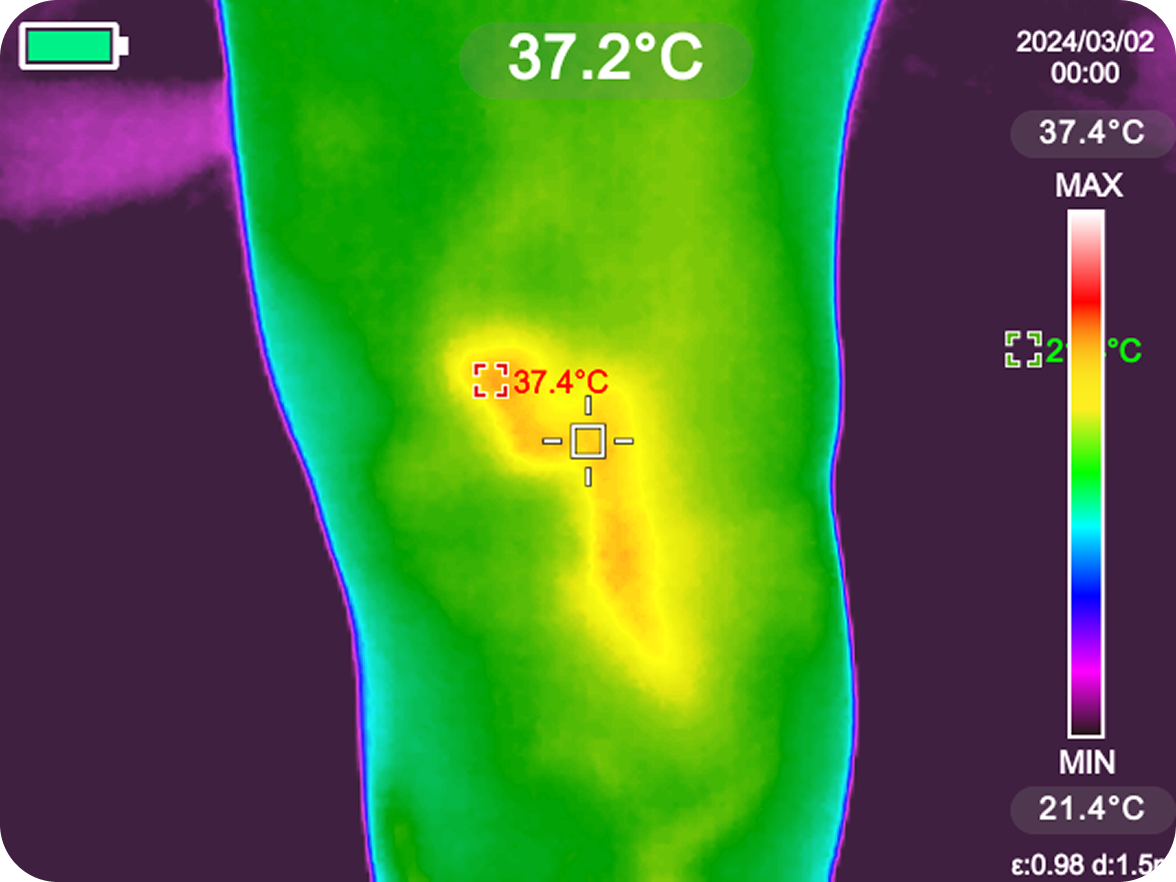
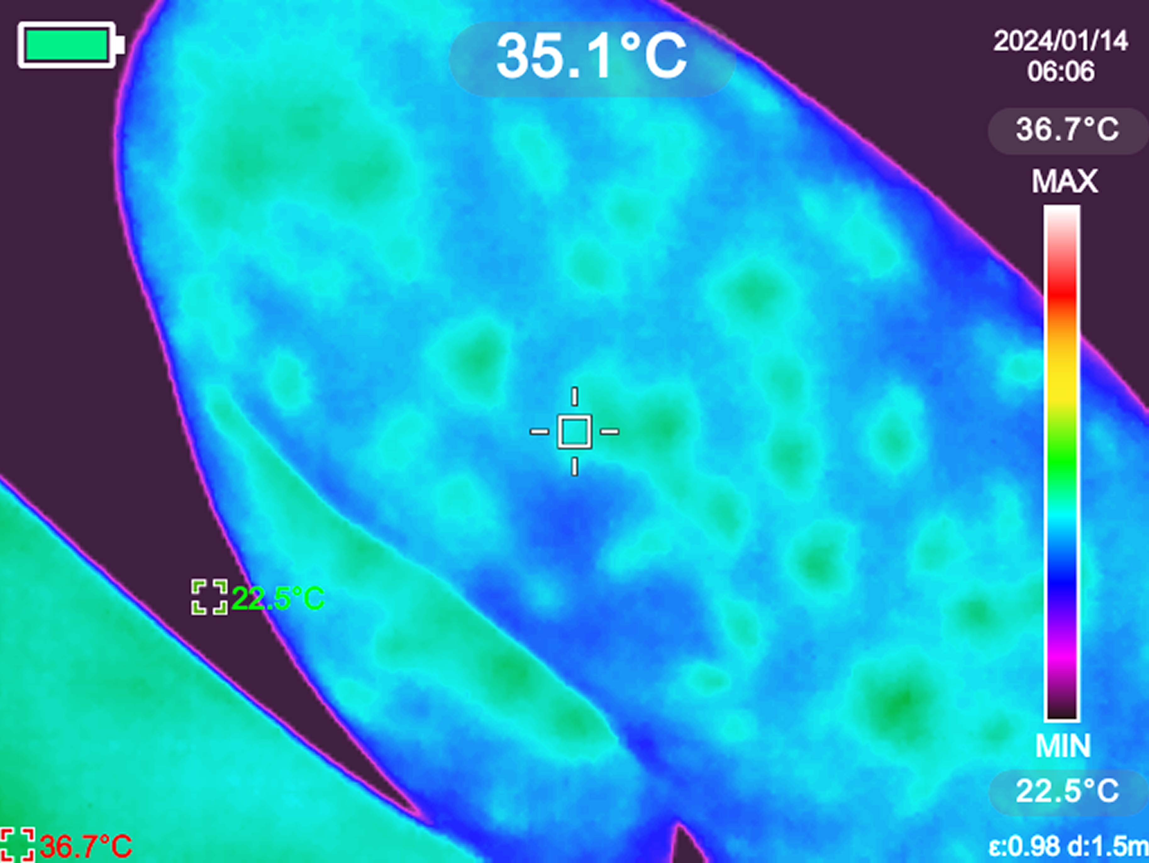
Thermography is a method of functional diagnostics based on the registration of infrared radiation from the human body. It operates by detecting thermal asymmetry in adjacent areas of the body.
The method is harmless (non-invasive), as the body is not subjected to radiation or any damage.

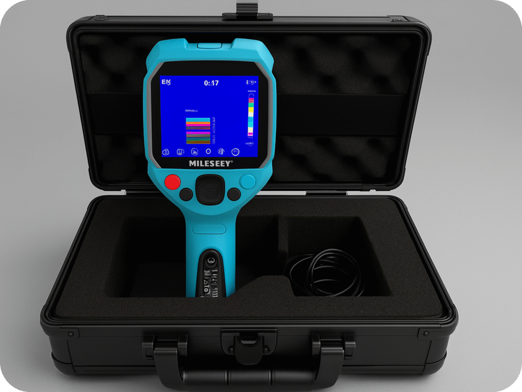
The possibility of detecting disease using thermograms is based on the fact that various pathological conditions affect the distribution and intensity of thermal (infrared) radiation. Using the method of thermography, it is possible to perform topical diagnosis of tumor foci, conduct differential diagnosis of benign and malignant neoplasms, and predict the course of the disease.
This method is most often used for the diagnosis of tumors of the mammary gland, skin, and soft tissues. The physiological basis of thermography is the increase in the intensity of infrared radiation over the pathological focus due to the enhancement of its blood supply and metabolic processes, or the decrease in its intensity in areas with reduced regional blood supply and accompanying changes in the surrounding tissues.
Thermography registers not structures, but physical processes occurring in the body. Therefore, it helps to detect the process of neoplasm at an initial stage, when the physical body of the tumor is extremely small in size. It is known that the normal body surface temperature provides a characteristic thermographic image. The lowest temperature is found in the distal parts of the limbs, the tip of the nose, and the auricles. The highest temperature is in the areas of the lips, cheeks, neck, and armpits.
For other body areas, the temperature is more moderate and constant. Diurnal fluctuations in skin temperature can reach 0,3-0,4 °С. It should be noted that thermoasymmetry is not always an absolute sign of pathology. Physiological thermoasymmetry exists, where temperature differences are minor and differ from pathological ones. However, it is generally accepted that the physiological temperature difference between symmetrical zones does not exceed 0,2-0,4 °С.
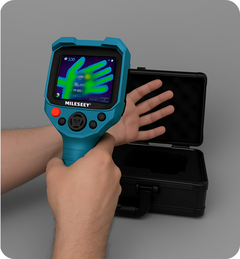
Areas of varicose veins are clearly visible in the images due to increased blood filling, and lymphadenopathy is visible due to locally inflammatory and congested phenomena in the lymph nodes.